National Center for Geriatrics and
DMA homeIntroduction Research Group GroupsGroupsAlbumNotice Board
    Department
    Department

Michan Group

    Department

Yamakoshi Group

    Department

Naoe Group

    Department
    Department

Michan Group Page

Back to Japanese Page

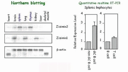
3. Research Projects

Our research style is aiming for a multiple fields spread over gerontology and immunology using a temporal or spacial gene targeting with Cre-loxP system. Our current experimental projects based on murine embryonic technology in addition to molecular biology and immunology, furthermore, gene and regenerative technology of embryonic stem cells are introducred below. We are preceding all these projects keeping originality and intelligibility.

A. Molecular analysis of a weakness of immune response along with aging in acquired immune system.

A weakness of acquired immune response with aging is closely related with a trigger of many diseases in the aged. Especially, it is important to clarify the machinery of induction and maintenance of immunological memory that can protect our body from the second invasion caused by the various infectious diseases. In other words, consequently, this research expected for making a progress in immune therapies with vaccine or an antibody is useful for materializing a sound aged society.

We proceed with the molecular research in the functional cells of the acquired immune response, such as B cells, T cells or dendrite cells, and establish the experimental animal model to analyze the weakness of aged immune response in a body. Ultimately, we aim to offer the immune system to be overcome and restored to human being. Furthermore, we also research to screen the functional biomarkers for monitoring the process of decline in the immune respons

<Our results in this part>

Zizimin 2 is a novel, DOCK180-related Cdc42 guanine nucleotide exchange factor expressed predominantly in lymphocytes.

Fig.2 We identified a novel CDM family molecule, zizimin2, as a functional molecule highly expressed in splenic germinal center by a suppression subtraction hybridization from C57Bl/6 mice immunized NP-CGG.(Nishikimi et.al, 2005)


Fig.3 We revealed that zizimin2 was capable to bind and activate cdc42 guanine nucleotide exchange factor expressed predominantly in lymphocytes (Nishikimi et.al, 2005)


B. Functional analysis of cellular senescence related genes in aging and research on a

treatment for their linked diseases

It is an urgent problem to construct the sound aged society in which the aged have lived their useful life. This project aims to analyze the molecular mechanism of fundamental regulation of aging on the basis of that of cellular senescence, that is, the growth arrest of the cells.

In the concrete, we focus to screen the senescent specific highly expressed genes from mouse embryonic fibroblasts (MEFs) and study their physiological functions after suffering the various stresses such as an oxidative stress. And consequently, we aim to throw a new light upon the common basic mechanism of aging in molecular, cellular and even a whole body level. We also investigate the phenotype of the model mice derived from our interest gene related-diseases and try to find a breakthrough to overcome them.


*<Our results in this part>

In a known inflamental cytokine, IL-1 gene family, especially, IL-1ƒÀ (ligand) and IL-1Ra (receptor antagonist) were involved in MEF cellular senescence through a p38-mediated signal pathway.

Fig.4. Schematic representation of a signaling pathway involved by IL-1 gene family in a stress-induced cellular senescence. (Premature senescence)


Fig.5. We examined the growth rate of MEFs from IL-1Ra deficient mice and revealed its distinguishable accelerated senescence in growth curve (A) and BrdU incorporation (B) comparing to wildtype MEFs culture. (Uekawa et.al 2004)


TARSH (Target of Nesh), originally identified as a novel cellular senescence related gene, represents a lung-specific gene expression. (Fig.6).We note that TARSH is a functional cellular senescence related gene involving in tumor suppression, since its mRNA expression was markedly declined in lung cancer cell lines that we have tested.


Fig.6 Tissue distribution of mTARSH mRNA expression. Poly(A)+ RNAs from 4-week-old C57Bl/6J mice tissues were analyzed by Northern blotting (left) and( Quantitative RT-PCR analysis was performed using total RNA from lung tissue of 1-, 8-, and 20-month-old C57Bl/6J mice. (Uekawa et.al 2005j

Fig.7 RT-PCR analysis of lung cancer cell lines (LCMS, A549, LCP, and LK-2) and normal lung cell lines (CCD-14Br and MRC-5) was performed at the linear range of cycle numbers (upper). Quantitative realtime RT-PCR analysis was performed using the same cell lines above. The bars represent the relative hTARSH mRNA level in CCD-14Br after normalization to ƒÀ-actin.
(Uekawa et.al 2005j

Michan Group Top Page

Pubulicatgions

DMA Home | Introducion| Research

item2
DMA homeIntroduction Research Group GroupsGroupsAlbumNotice Board